
When the eye needed to come closer
Microsurgery was not born in a single moment.
It took shape in stages, across many specialties, as surgery began to be performed under the microscope, with dedicated instruments and sutures for very small blood vessels, nerves and lymphatic vessels [1].
But before a small vessel could be joined, there was a more basic question.
How do you see it clearly enough?
In 1921, the Swedish otolaryngologist Carl Olof Nylén used a monocular microscope in ear surgery. According to Mudry’s review, this was the first use of the microscope in ear surgery [2].
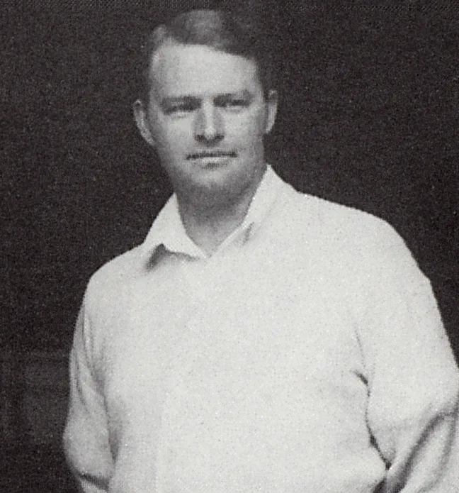
Just a year later, Gunnar Holmgren replaced the monocular microscope with a binocular one [2].
A change that seemed small.
But the road ahead was still long.
Microscopes of that time had a narrow field of view and weak light. Only in 1951, when the model perfected by Littmann and Zeiss appeared, did the operating microscope gradually come into wide use [2].
Surgeons could now look deeper into a very small world.
What would come next?
When small vessels could be joined again
In 1960, Julius H. Jacobson II and Ernesto L. Suarez reported a technique for joining small blood vessels under the microscope [3]. Two years later, they published further work on microvascular surgery [4].
The microscope no longer only helped the surgeon see.
It became part of the operation itself.
In 1966, Harry J. Buncke and W.P. Schulz reported the complete replantation of a rabbit ear by joining very small vessels [5]. It was a step towards operations on people.
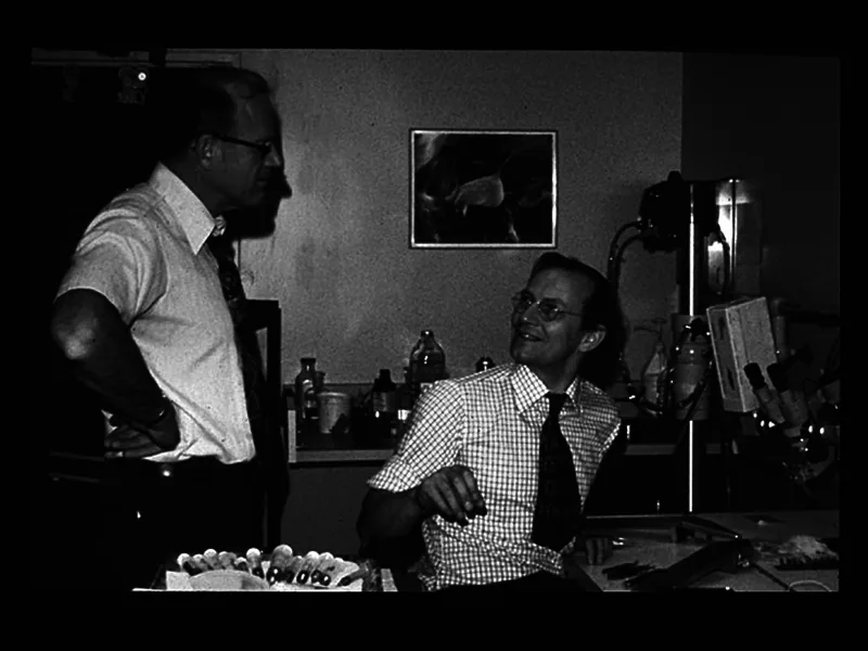
And in Nara, Japan, that step had already been taken.
In 1965, Shigeo Komatsu and Susumu Tamai successfully replanted a completely amputated thumb; the result was published in 1968 [6].
Structures once too small for conventional surgery were now within the surgeon’s reach.
From the hand to the brain
Microsurgery went further.
Not only in peripheral vessels.
In 1967, M. Gazi Yaşargil performed the first superficial temporal artery to middle cerebral artery (STA–MCA) bypass [7]. In 1968, he and R.M.P. Donaghy published on microvascular surgical technique [8].
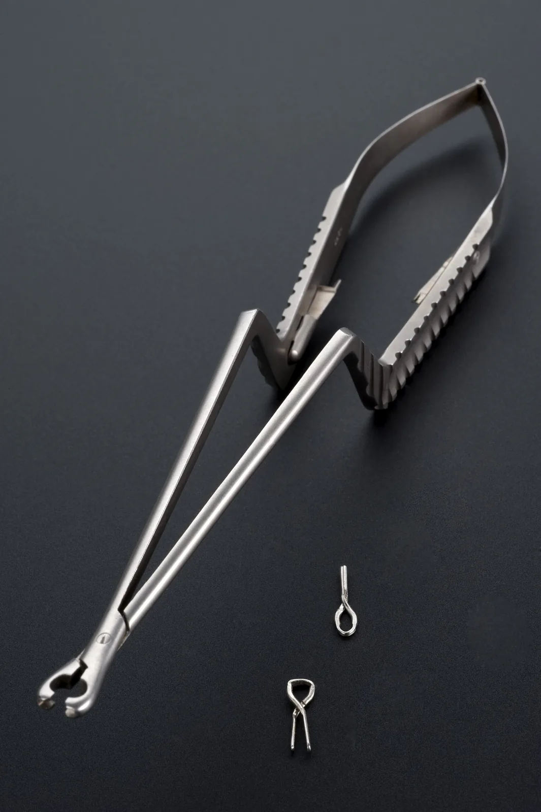
Once small vessels could be joined, another question followed.
If a vessel can be joined, could a whole piece of tissue be moved somewhere else?
In 1973, Rollin K. Daniel and G. Ian Taylor published the transfer of an island flap to a distant site by microvascular anastomosis [9]. In 1974, Kiyonori Harii and colleagues reported free transfer of hair-bearing scalp flaps [10].
Microsurgery was no longer the story of a single manoeuvre.
It opened a new way to move and rebuild tissue.
When every detail of an instrument mattered
Seeing small structures was not enough.
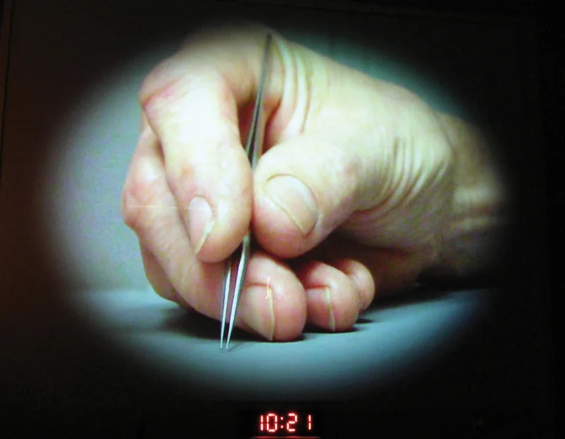
The hands also needed instruments made for that scale.
In 1974, Robert D. Acland described a new vessel clamp and a device for holding stay sutures in microvascular anastomosis [11].
A technical detail.
Yet it showed something important: as the structures grow smaller, the instruments must change with them.
That is why microsurgery has always rested on three things together: the operating microscope, microsurgical instruments and fine sutures [1].
Without any one of them, the hands cannot go further into that very small world.
And the limit grew smaller once more
In 1989, Isao Koshima and S. Soeda described a skin flap based on the inferior epigastric artery without the rectus abdominis muscle, laying the ground for perforator flaps [12].
The journey towards the smaller did not stop there.
In 2000, Koshima and colleagues reported supermicrosurgical lymphaticovenular anastomosis for upper-limb lymphoedema [13].
From a monocular microscope in ear surgery in 1921 to lymphaticovenular anastomoses, microsurgery travelled for nearly eight decades.
Not in one leap.
But in many small steps.
A microscope that saw more clearly. A technique for smaller vessels. An instrument that fitted the hand better. A finer suture.
Each time a limit seemed fixed, surgical hands moved a little further.
Today, microsurgery is part of plastic, hand, neuro- and ENT surgery.
Yet behind it all lies something very simple:
To work on the smallest structures, you must first be able to see them.
References
- Tamai S. History of microsurgery. Plast Reconstr Surg. 2009;124(6 Suppl):e282-e294. doi.org/10.1097/PRS.0b013e3181bf825e
- Mudry A. The history of the microscope for use in ear surgery. Am J Otol. 2000;21(6):877-86. pubmed.ncbi.nlm.nih.gov/11078079/
- Jacobson JH, Suarez EL. Microsurgery in anastomosis of small vessels. Surg Forum. 1960;11:243-5.
- Jacobson JH 2nd, Suarez EL. Microvascular surgery. Dis Chest. 1962;41:220-4. doi.org/10.1378/chest.41.2.220
- Buncke HJ Jr, Schulz WP. Total ear reimplantation in the rabbit utilising microminiature vascular anastomoses. Br J Plast Surg. 1966;19(1):15-22. doi.org/10.1016/s0007-1226(66)80003-6
- Komatsu S, Tamai S. Successful replantation of a completely cut-off thumb. Plast Reconstr Surg. 1968;42(4):374-7. doi.org/10.1097/00006534-196810000-00021
- Vilela MD, Newell DW. Superficial temporal artery to middle cerebral artery bypass: past, present, and future. Neurosurg Focus. 2008;24(2):E2. doi.org/10.3171/FOC/2008/24/2/E2
- Donaghy RM, Yasargil G. Microangeional surgery and its techniques. Prog Brain Res. 1968;30:263-7. doi.org/10.1016/s0079-6123(08)61469-7
- Daniel RK, Taylor GI. Distant transfer of an island flap by microvascular anastomoses. Plast Reconstr Surg. 1973;52(2):111-7. doi.org/10.1097/00006534-197308000-00001
- Harii K, Omori K, Omori S. Hair transplantation with free scalp flaps. Plast Reconstr Surg. 1974;53(4):410-3. doi.org/10.1097/00006534-197404000-00005
- Acland RD. Microvascular anastomosis: a device for holding stay sutures and a new vascular clamp. Surgery. 1974;75(2):185-7. pubmed.ncbi.nlm.nih.gov/4590759/
- Koshima I, Soeda S. Inferior epigastric artery skin flaps without rectus abdominis muscle. Br J Plast Surg. 1989;42(6):645-8. doi.org/10.1016/0007-1226(89)90075-1
- Koshima I, Inagawa K, Urushibara K, Moriguchi T. Supermicrosurgical lymphaticovenular anastomosis for the treatment of lymphedema in the upper extremities. J Reconstr Microsurg. 2000;16(6):437-42. doi.org/10.1055/s-2006-947150