
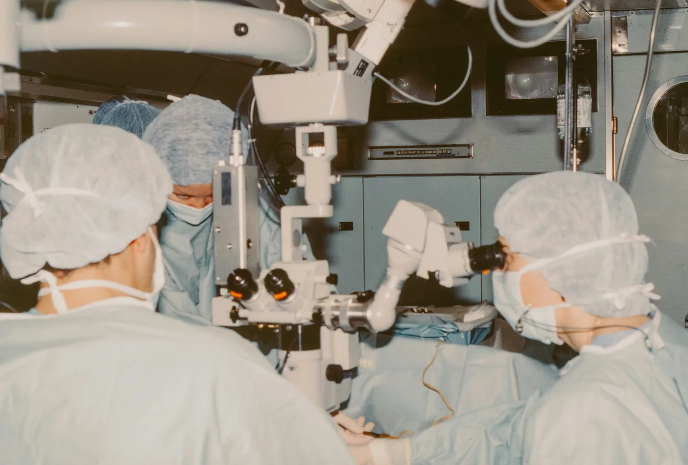
When one eye entered the operating room
The story of the operating microscope does not begin with a large system.
It begins with a single eyepiece.
In 1921, in Stockholm, Sweden, the otolaryngologist Carl Olof Nylén used a monocular microscope in ear surgery on a patient with chronic otitis. According to Mudry, this was the first use of the microscope in ear surgery [2, 3].
But seeing larger did not yet mean seeing better.
Early microscopes had a narrow field of view, a short working distance, weak light and poor stability, so at first they were rarely used [2].
Just a year later, Gunnar Holmgren, from the same department as Nylén, moved to a binocular microscope with an attached light source.
Two eyepieces gave depth perception. The light kept the image from darkening under magnification [2, 5].
The microscope now had a second eye, and light.
But to truly belong in the operating room, it still had to learn to stand firm.
When the microscope learned to stand
In 1938, Tullio and Calicetti at the University of Parma built a heavy counterweighted stand and added a prism so that an assistant could view the same field [5].
The microscope was no longer for one person only.
In 1946, Richard A. Perritt brought the binocular microscope into ophthalmic surgery [5].
Then, in 1953, a major step arrived.
Zeiss introduced the OPMI 1, designed by Hans Littmann: magnification could change without changing focus, illumination was coaxial, and the stand had a swivel arm based on an idea from Horst Wullstein [1, 5, 6].
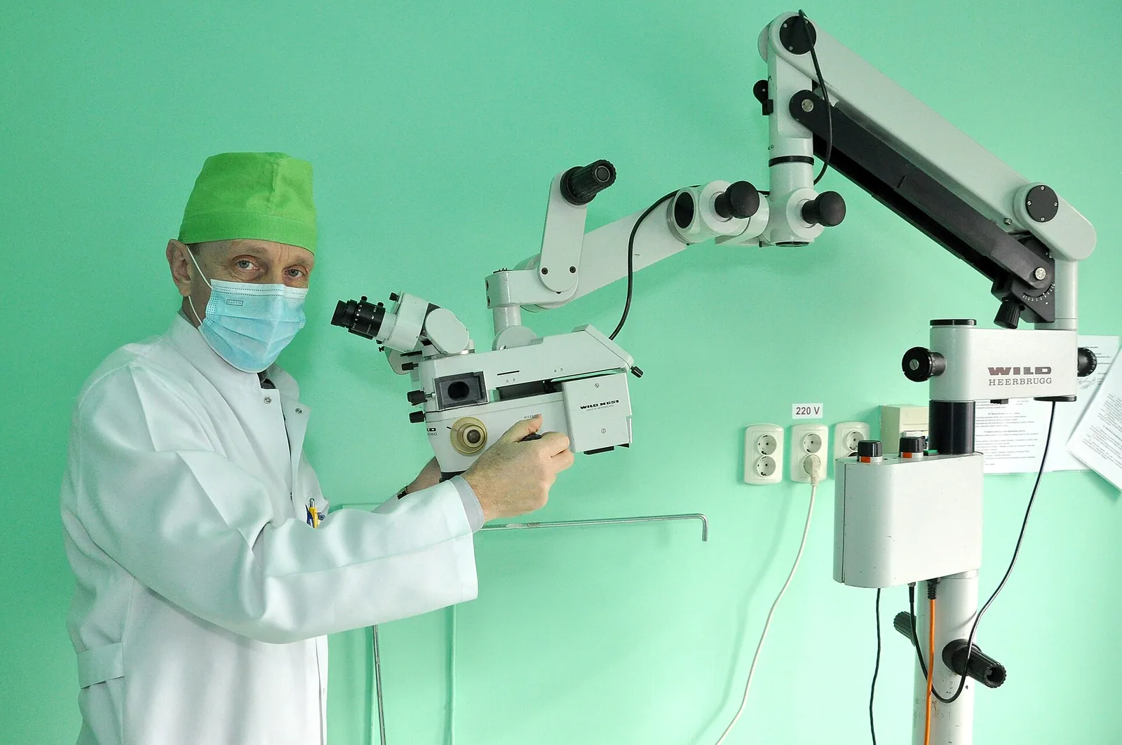
Three years later, Joaquín Barraquer designed a ceiling-mounted suspension [5].
The optics changed.
The light changed.
The way the microscope stood beside the surgeon changed too.
And then it moved into other surgical fields.
When the microscope reached the brain and the vessels
In 1957, Theodore Kurze brought the microscope into neurosurgery, removing a facial nerve schwannoma in a five-year-old child [1, 5].
In 1960, Julius Jacobson successfully performed a microvascular anastomosis; according to the literature, this was the first application of the technique to small vessels [6].
The microscope no longer only magnified anatomy.
It accompanied work on ever smaller structures.
In 1964, Littmann’s Diploscope beam splitter allowed two surgeons to observe together [5]. In 1965, Pool and Colton used the microscope in intracranial aneurysm surgery [11].
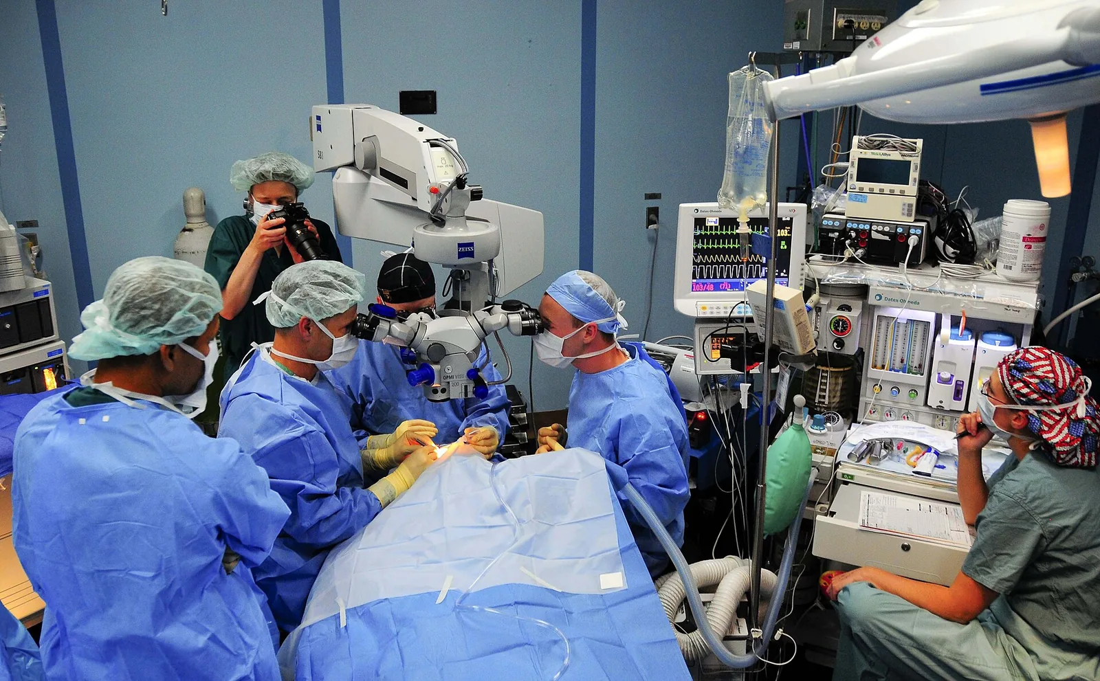
In Zürich, M. G. Yaşargil worked with the company Contraves to refine a balanced stand based on a parallelogram arm, adding illumination, video and photography [7].
After training in the department of R. M. P. Donaghy in Vermont, USA, microneurosurgery became routine in Zürich from 1967 [7, 8].
From the ear and the eye to the nerves and the vessels.
But the story did not stop at seeing structures.
Could the microscope also reveal what the eye cannot see?
When the image began to carry more information
In 2003, Raabe and colleagues reported near-infrared indocyanine green (ICG) videoangiography through the microscope, assessing blood flow during surgery [9].
In 2006, the phase III randomised trial by Stummer and colleagues evaluated 5-ALA fluorescence-guided surgery for malignant glioma [10].
After decades of optical and mechanical refinement, the image through the microscope could now integrate other ways of seeing.
From magnification to illumination.
From recording to fluorescence.
Each layer of technology added another way to look at the same field.
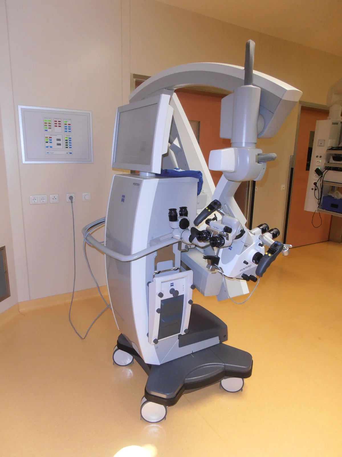
When the eyes left the eyepiece
Recently, another way of viewing has entered the operating room.
The 3D-4K exoscope shows the surgical field on a large screen, viewed through 3D glasses [11].
That change has brought differing observations. Some groups report benefits for working posture; others report limitations in depth of field [11].
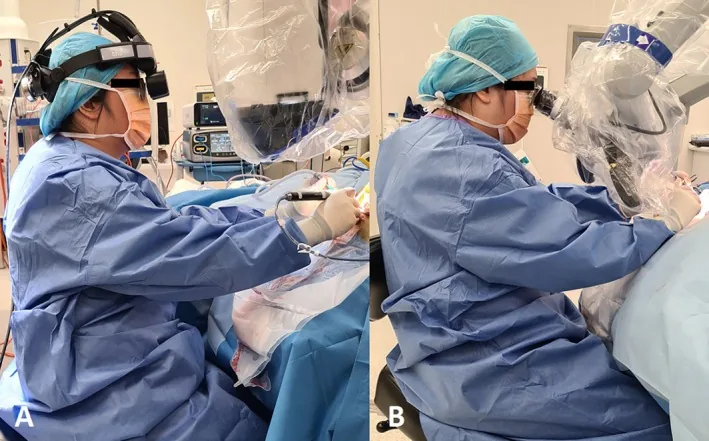
It is not the first time the surgeon’s way of seeing has changed.
From one eye to two. From weak light to coaxial illumination. From one observer to two surgeons looking together. From optical images to video, photography and integrated imaging.
Over more than a century, the operating microscope has been refined step by step in optics, illumination, stands, recording and integrated imaging. Each step is tied to surgeons who used it in the operating room [1, 4].
The microscope of 1921 has come a very long way.
Yet the need remains close to where it began:
When the hands work on ever smaller structures, the eyes need to see more.
References
- Uluç K, Kujoth GC, Başkaya MK. Operating microscopes: past, present, and future. Neurosurg Focus. 2009;27(3):E4. doi.org/10.3171/2009.6.FOCUS09120
- Mudry A. The history of the microscope for use in ear surgery. Am J Otol. 2000;21(6):877-86. pubmed.ncbi.nlm.nih.gov/11078079/
- Nylén CO. The microscope in aural surgery, its first use and later development. Acta Otolaryngol. 1954;43(Suppl 116):226-40. doi.org/10.3109/00016485409130299
- Kriss TC, Kriss VM. History of the operating microscope: from magnifying glass to microneurosurgery. Neurosurgery. 1998;42(4):899-907. doi.org/10.1097/00006123-199804000-00116
- Ma L, Fei B. Comprehensive review of surgical microscopes: technology development and medical applications. J Biomed Opt. 2021;26(1):010901. doi.org/10.1117/1.JBO.26.1.010901
- Moritz WR, et al. The history and innovations of blood vessel anastomosis. Bioengineering (Basel). 2022;9(2):75. doi.org/10.3390/bioengineering9020075
- Stienen MN, et al. UniversitätsSpital Zürich: 80 years of neurosurgical patient care in Switzerland. Acta Neurochir (Wien). 2018;160(1):3-22. doi.org/10.1007/s00701-017-3357-z
- Yaşargil MG. Personal considerations on the history of microneurosurgery. J Neurosurg. 2010;112(6):1163-75. doi.org/10.3171/2009.7.JNS091124
- Raabe A, et al. Near-infrared indocyanine green video angiography: a new method for intraoperative assessment of vascular flow. Neurosurgery. 2003;52(1):132-9. doi.org/10.1097/00006123-200301000-00017
- Stummer W, et al. Fluorescence-guided surgery with 5-aminolevulinic acid for resection of malignant glioma: a randomised controlled multicentre phase III trial. Lancet Oncol. 2006;7(5):392-401. doi.org/10.1016/S1470-2045(06)70665-9
- Oertel J, Keiner D. Novel devices for intraoperative visualization in neurosurgical procedures: current state and prospect of using the exoscope. Acta Neurochir (Wien). 2021;163(8):2117-9. doi.org/10.1007/s00701-021-04839-7